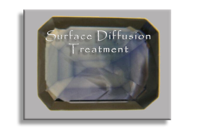
New research shows that the titanium surface diffusion treatment is being polished off the facets, but left inside cavities and fractures.
Stone Group Laboratories
Being aware that every blue sapphire might be surface diffused can save you from a costly mistake. But now there is something even more to consider.
To find evidence for conventional titanium surface diffusion of blue sapphires you want to look for color
To see the treatment, it is best to make observations under a microscope using a light diffuser and/or liquid immersion.
Up until recently one would simply look for dark blue facet junctions, as there is more concentration of the titanium at the junction of two facets. You can see this easily in figure #1. Here we observe facet junctions that have a darker color due to the smaller surface area (both sides of a facet junction).
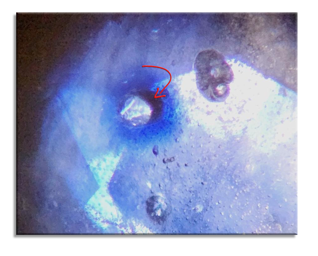
Now, look at the dark spot at around the 1 o’Clock position. What is going on there? (red arrow)
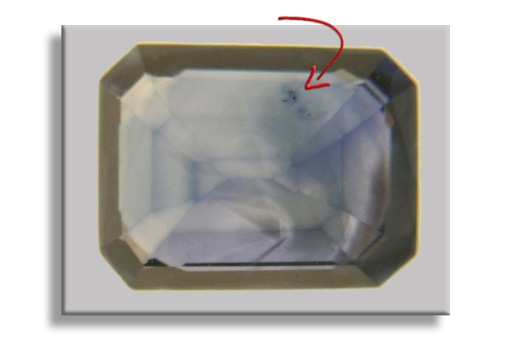
That small spot is where the titanium diffusion entered a surface breaking fissure and colored its edges.
Penetrating Fractures and Cavities
This added a new twist to this type of treatment because the evidence you have seen in figure 1 can be polished off. In Fig. 2a, the re-polished sapphire has effectively removed the expected color concentrations used as evidence.
Now more careful observations must be taken to look for other signs of possible color enhancement by cavity or “titanium fissure diffusion”. This has been observed as early as 1990, but the re-polishing concept is now being used to hide evidence.
Figure 2a, Evidence of surface diffusion has been removed
“Dark concentrations, or ‘bleeding,’ of color are often seen in and around surface reaching breaks and cavities in blue diffusion treated sapphires “Identification of Blue Diffusion-Treated Sapphires, Kane, et al. G&G Summer, 1990
Extra effort and closer observations need to be employed to catch any tell-tale signs. (Fig 2b) A very slight hint of the treatment is seen where the re-polish did not completely remove evidence.
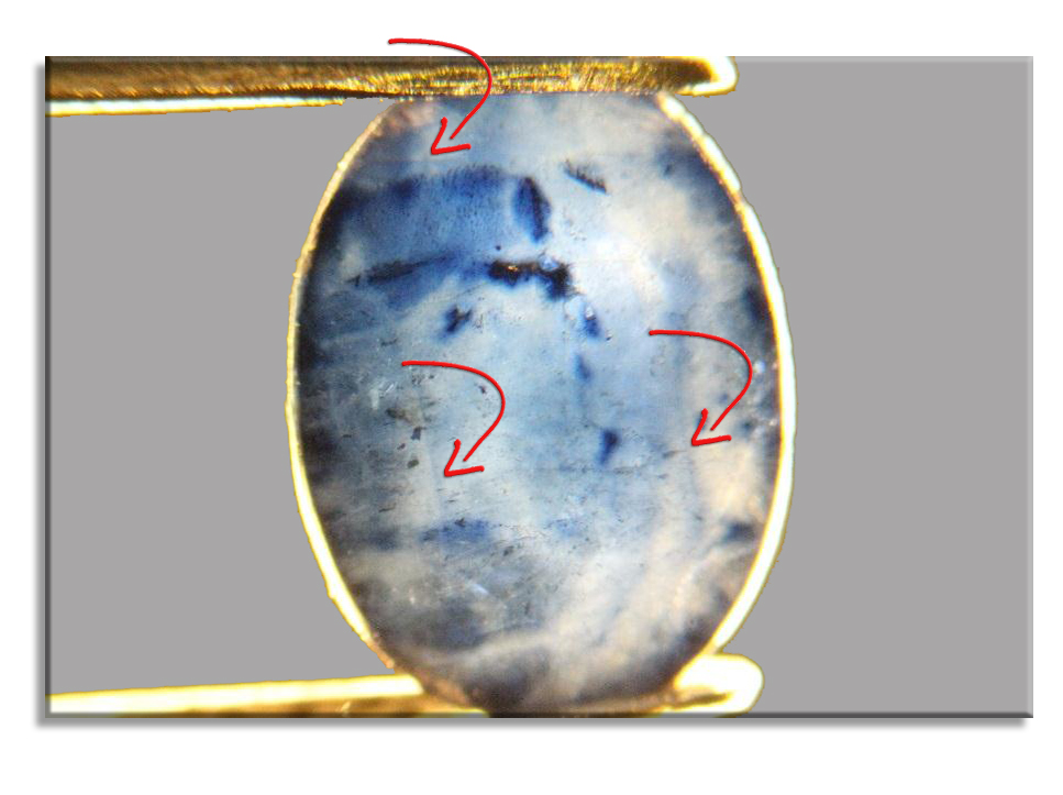
Look for the surface breaks or any minor cavity that can allow the titanium to reach even further into internal fissures.
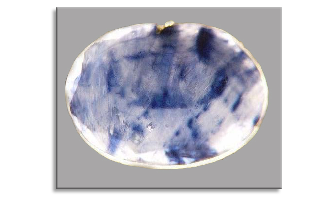
Figure 3 was taken at 35X. It shows a pit approximately 0.1 mm across. It has left clear evidence of the treatment as it colored the walls of this cavity. follow for more Roskingemnewsreport
Tap here to visit Stone Group Laboratories


