


This time, Billie Hughes gives us something rarely seen: a before-and-after-and-after-again look at what happens to an inclusion during heat treatment.
As gemologists, we are always looking for clues of enhancement. In this case, we have a natural Burma ruby, so one thing we would be looking for is evidence of heat treatment.
To help the rest of us, Billie Hughes shares a Burma ruby containing a spinel crystal inclusion. The ruby was photographed before heating, after low-temperature heating, and then again after heating to a much higher temperature.
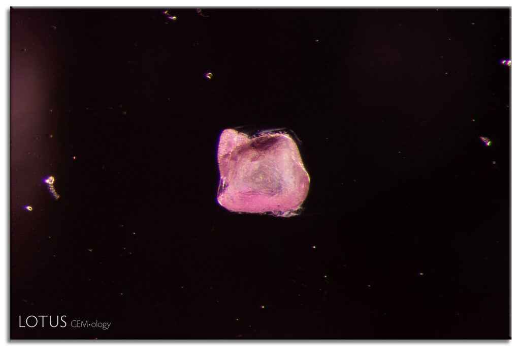
Here is the crystal in its original, unheated state.
A: (Unheated) A transparent spinel crystal in an unheated Burmese ruby.

Photomicrograph by E. Billie Hughes; field of view 1.5 mm.
Then the ruby was heated to a relatively low temperature of 1100°C.
The changes to the spinel crystal are obvious here, but would we be so confident in calling heat treatment if we didn’t have the before image for comparison? Take a close look at this shot.
B: (1100°C) When the ruby was heated to 1100°C, the spinel crystal began to change in appearance, developing elongated lines along its surface.
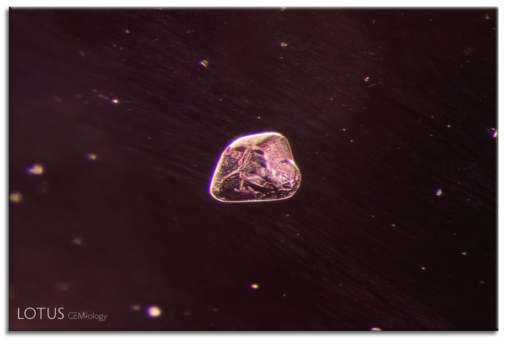
Photomicrograph by E. Billie Hughes; field of view 1.5 mm.
The ruby was then heated a second time, this time to 1500°C.
The result is dramatic.
This is the third shot.
C: (1500°C) After heating to 1500°C, the crystal took on a frosty, whitish appearance.
Photomicrograph by E. Billie Hughes; field of view 1.5 mm.
As impressive as the transformation of the spinel crystal is, another challenge is hidden in these images. After each heating experiment, the ruby had to be carefully re-oriented in the microscope so the same tiny spinel crystal could be photographed from virtually the same position.
“Each specimen was examined and photographed in the microscope before and after every stage of treatment. The changes to solid inclusions tended to follow a pattern as the samples were heated to increasing temperatures. Notable changes included the following:”
- Alterations to fissure stains
- Formation and subsequent healing of fissures
and, what we see here in these images, 3. Alterations in the appearance of solid mineral inclusions.
“Many crystals began to display changes in opacity and texture as they were heated to progressively higher temperatures. Their tendency was to become less transparent, often developing a white interior. In many cases they also developed a more textured exterior, sometimes becoming so ‘frosted’ that they appeared opaque.”
“These observations were not limited to spinel inclusions but were seen across a variety of solid inclusions. Similar patterns were noted with mica, calcite, sphene, iron sulfide, diaspore, primary rutile, scapolite, feldspar, and apatite.”
◈
An Added Bonus! Raman Spectra
Billie points out that “the Raman spectra of spinel inclusions can also alter after heat treatment (particularly with the broadening of the peak around 405–120 cm-1). We have a nice example of this in Figure 29 in the same article. (Note that it’s not the same exact crystal as the one pictured here, or in Figure 16 in the article.)”
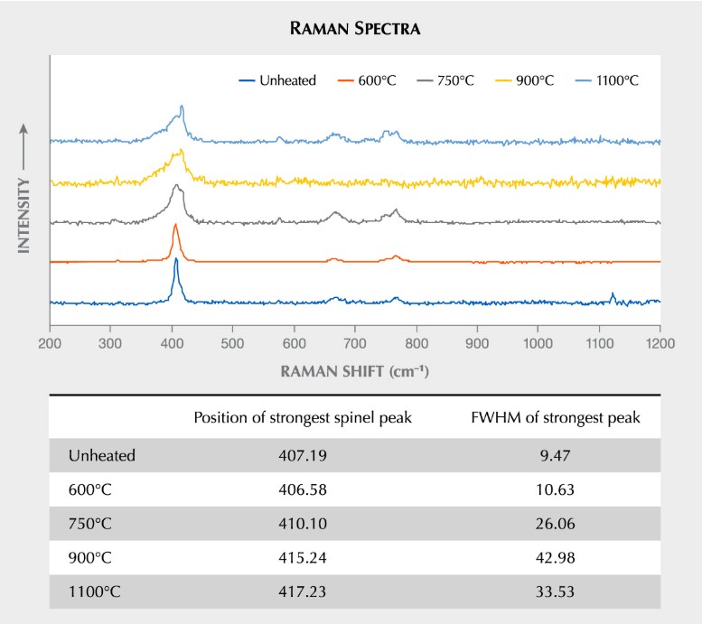
From Lotus Gemology – Canary in a Ruby Mine
Canary in the Ruby Mine
All of these images can be seen in Lotus’ research paper: Canary in the Ruby Mine — Low-Temperature Heating of Burmese Ruby.
https://lotusgemology.com/resources/articles/501-canary-in-the-ruby-mine-low-temperature-heating-of-burmese-ruby
( and G&G – https://www.gia.edu/gems-gemology/winter-2022-burmese-ruby)
E. Billie Hughes, FGA, is a co-founder of Bangkok-based Lotus Gemology, a respected gemological laboratory and educational resource.
Tap Here see more about this and other inclusion images at LotusGemology.com!
… and Tap Here for more of Billie Hughes’ images right here!